AEC gives due Importance to Modern Medical Devices in your Eye care
At Acuity Eye Centre we provide a great range of modern technologies to best examine your eyes for eye disease, binocular vision problems, and refractive problems (for glasses and/or contact lenses).
The equipment used by our front staff in our pre-testing room is computerised and does the following:
- The autorefractor measures the curvature of your corneas (important in fitting contact lenses and sometimes in diagnosing corneal disease).
- The autorefractor also estimates your refractive error (an objective estimation of what your eyeglass prescription may be). Your doctor of optometry requires another refractive testing (both objective and subjective) before a final prescription can be written.
- Nidek Digital Lensometer measures the refractive power of the glasses you are already using
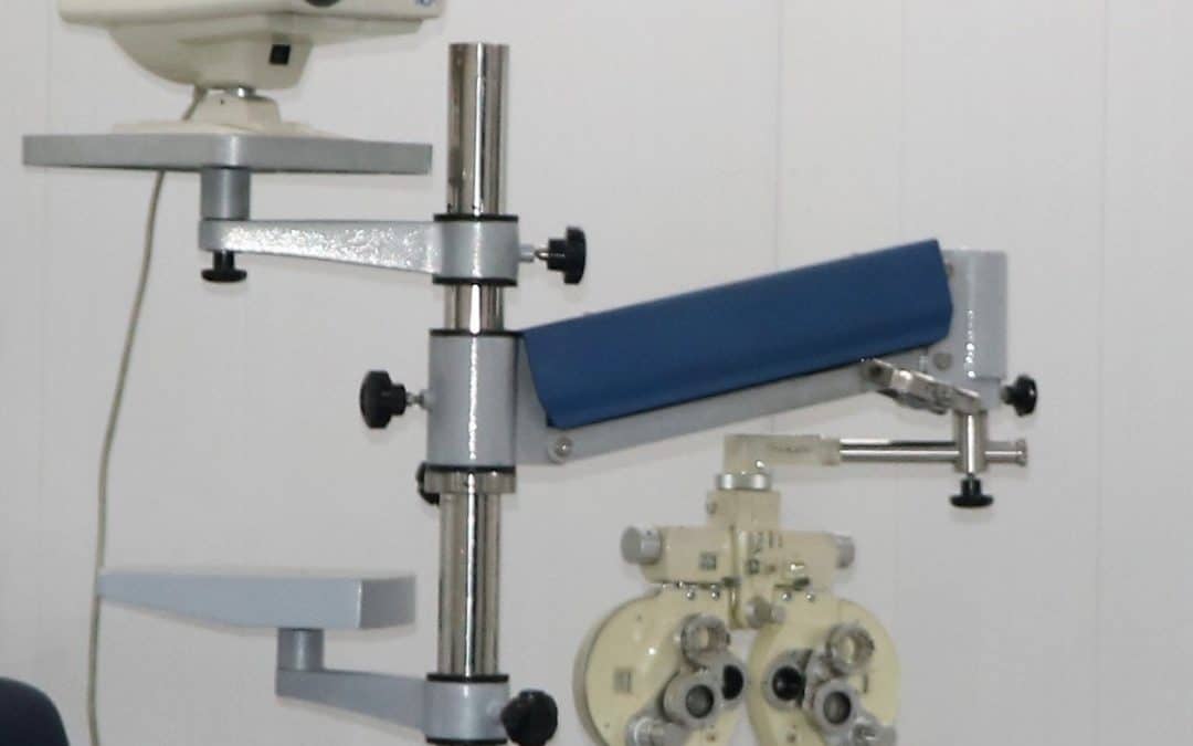
The Phoroptor

In the doctor’s examination room, he utilises a variety of specialised equipment. The phoroptor is an instrument that greatly aids in the determination of your spectacle prescription. The microscope (slit-lamp biomicroscope) is used to assess how healthy your eyes are (both inside and out). There are also handheld and headset instruments which allow for a thorough evaluation of back of the eyes (retina). Dilation with eye drops is often required.
Visual Field Testing

Visual field testing is an automated, computerised instrument that tests the sensitivity of various points in your peripheral vision. This is useful when looking for glaucoma, pituitary tumours, visual field loss from strokes and many other situations.
OCT

OCT is a tool to aid in the diagnosis and management of retinal disease (such as age-related macular degeneration, cystoid macular oedema, macular holes) and glaucoma. It uses light to scan the retina and optic disc to give a cross-sectional (that is to say three dimensional) image of those regions.” It allows more accurate and faster diagnosis of these conditions which ultimately means better care for the patient.
It is not used for peripheral retinal problems such as retinal detachments.
Additionally, there are other tests/procedures the doctor may recommend after the eye exam. These include visual field testing, Optical Coherence Tomography (OCT) and pachymetery.
Digital Slit Lamp Photography

Slit lamp photography to practice can offer several advantages. The fine detail can allow detection of slight changes of corneal findings, such as resolving corneal infections or edema. These recordings can also enhance patient care when a patient is being followed by multiple physicians for the same condition. By projecting the images to a larger screen, these photos can also serve as excellent teaching tools for patients to explain everything from meibomian gland disease to cataracts.
Contact Our Team:
If you are looking for any of below services, please fill the form below, one of our team member will get in to provide you with full facilitation:
1- Comprehensive Primary Eye Exam/ Consultation
Consultation ::: Adult Eye Examination and Consultation
Consultation ::: Children Eye Examination Refraction Consultation
Consultation ::: Infant Eye Examination Refraction Consultation
2-Secondary Follow up Eye Examination and Consultations
Followup ::: Examination under Sedation for Kids (After Initial Consultation)
Followup ::: Dilated Fundus Examination(DFE)
Followup ::: Cycloplegic Refraction and DFE
3-Diagnostic Eye Test
Diagnostic ::: OCT
Diagnostic ::: Angio OCT
Diagnostic ::: Anterior Segment OCT
Diagnostic ::: Pachymetery
Diagnostic ::: Perimetery/Visual Fields
Diagnostic ::: Hess Chart/Digital Squint Assessment/Digital Diplopia Test
Diagnostic ::: Digital Colour vision test